
Structure reveals function—driving breakthroughs in biology, chemistry, and medicine.
Understanding molecular structure is key to understanding function. By revealing the precise arrangement of atoms, structural biology enables scientists to uncover how proteins and small molecules work, how diseases develop, and how new therapies can be designed. Continued advances in imaging and detection technologies are pushing the limits of resolution, allowing researchers to visualize biological machinery in unprecedented detail. As a manufacturer of high-performance direct detection cameras supporting these discoveries, we’re inspired by the breakthroughs made possible through structural imaging. Here, we highlight five landmark molecular structures whose extraordinary resolution has expanded the boundaries of science.
| Resolution | Structure | Technique | Detector | Authors | Radiation Source |
|---|---|---|---|---|---|
|
0.48 Å |
X-Ray Diffraction |
MAR CCD 165 mm |
Schmidt, A., Teeter, M., Weckert, E., Lamzin, V.S. |
PETRA II, DESY
BEAMLINE PETRA1 |
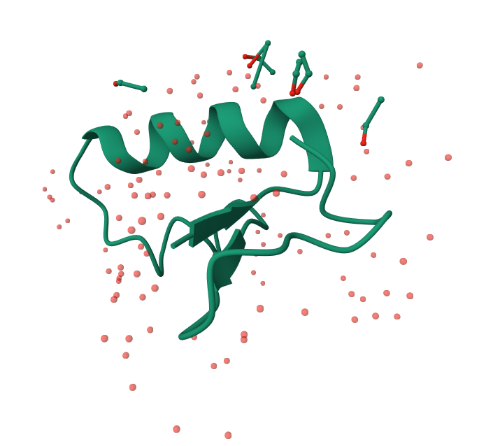
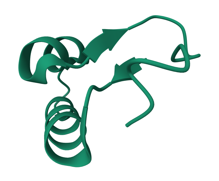
Figure 1: Small (4.92 kDa – 46 amino acids), rigid, highly ordered structure that diffracts to extremely high resolution makes it a good benchmark molecule. Biologically, crambin is a small seed protein from Crambe abyssinica that functions as a seed-storage and protective protein.
| Resolution | Structure | Technique | Detector | Authors | Radiation Source |
|---|---|---|---|---|---|
|
0.48 Å |
X-Ray Diffraction |
RAYONIX CCD
MX225HE |
Hirano, Y., Takeda, K., Miki, K., Lamzin, V.S. |
SPRING-8
BEAMLINE BL41XU |
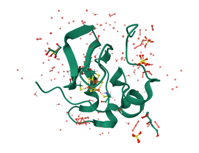
Figure 2: Ultra-high resolution (0.48 Å) X-ray structure of a high-potential iron–sulfur protein (HiPIP) with the distribution of valence electrons in addition to atom positions. Such features go beyond typical high resolution structures and reveal detailed electronic and bonding features in a biological electron-transfer center.
| Resolution | Structure | Technique | Detector | Authors | Radiation Source |
|---|---|---|---|---|---|
|
0.5 Å |
Micro-ED |
Direct Electron Apollo |
Takuma Fukumura; Takanori Nakane; Yuji Konyuba; Benjamin Bammes |
JEOL CryoARM 300 II |
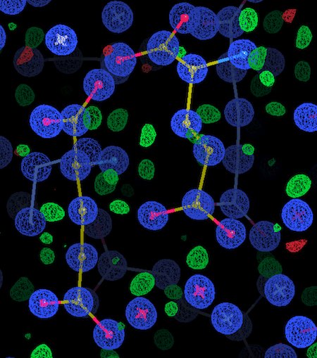
Figure 3: Positions of all atoms including hydrogens are visible in the ultra-high resolution (0.5 Å) electron density map. Significant as data was collected with an advanced MAPS detector (Apollo) using a conventional but powerful TEM (CryoARM300-2) rather than a synchrotron. Biologically, MSG is an important food additive/flavor enhancer and better understanding the structure can provide insight into taste receptors and other metabolic/neurological areas of research.
| Resolution | Structure | Technique | Detector | Authors | Radiation Source |
|---|---|---|---|---|---|
|
0.5 Å |
Micro-ED |
Direct Electron Apollo |
Takuma Fukumura; Takanori Nakane; Yuji Konyuba; Benjamin Bammes |
JEOL CryoARM 300 II |
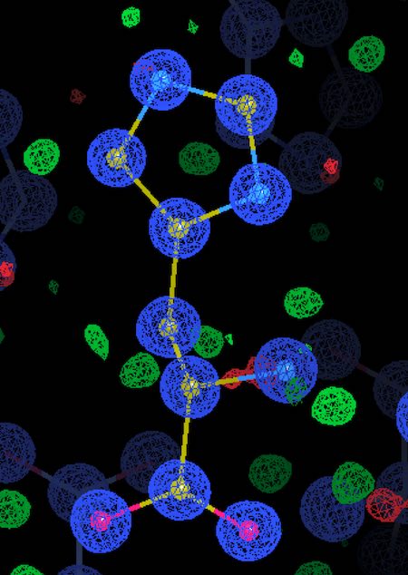
Figure 4: Strong low-frequency reflections require high counting rates to avoid coincidence loss but also high sensitivity to detect the very weak high-resolution reflections – Apollo handles both simultaneously. Improved resolution in histidine structures enables direct observation/placement of hydrogen atoms, unambiguous assignment of protonation and tautomeric states, and accurate electrostatic and bonding analysis.
| Resolution | Structure | Technique | Detector | Authors | Radiation Source |
|---|---|---|---|---|---|
|
0.54 Å |
X-Ray Diffraction |
Image Plate Mar Research |
Jelsch, C., Teeter, M.M., Lamzin, V., Pichon-Lesme, V., Blessing, B., Lecomte, C. |
EMBL/DESY, Hamburg Beamline BW7A |
Figure 5: Biologically, crambin is a small seed protein from Crambe abyssinica that functions as a seed-storage and protective protein.
Techniques
References:
