Electron Energy Loss Spectroscopy (EELS) is a transmission electron microscopy (TEM) technique in which electrons transmitted through a thin specimen (a few tens of nanometers thick) are dispersed by a spectrometer according to how much energy they have lost due to interactions with the specimen. EELS is a powerful technique that can allow microscopists to perform elemental analysis and also observe fine structure, and extract useful information about chemical bonding in their specimen.
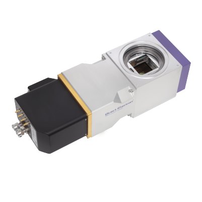
The recent development of the CEOS Energy Filtering and Imaging Device (CEFID) by CEOS GmbH [1] offers microscopists a choice of cameras from different manufacturers to use to collect EELS data. In a new video posted to the Direct Electron Youtube Channel, Dr. Benjamin Bammes, our Director of Research and Development makes the case for why the DE-16 Direct Detector is ideally suited to EELS experiments.
As illustrated in the video, a typical electron energy loss spectrum may be divided into three regions; a zero-loss peak, consisting of electrons that were transmitted through the specimen without losing a significant amount of energy; a low-loss region, containing information about features such as plasmons and band gap states; and finally a core-loss region, extending out to many keV of energy loss (not all of which can be shown in the video graphic), which contains information about the electron energy levels in the atoms that make up the specimen.
Ideally, an EELS detector would be able to acquire spectra that cover a wide energy range, containing the zero-loss peak as well as low-loss and core-loss information, while maintaining a high energy resolution so that fine structure can be clearly resolved.
Direct Electron’s patent pending HDR counting technique, which was described in a recent article on 4D STEM in Microscopy and Analysis [2], can allow us to detect single electron events in areas of the spectrum where the signal is relatively weak, without losing information from more intense parts of the spectrum, such as the zero-loss peak. Combined with a pixel count of 4096 in the energy dispersive direction, HDR counting can allow the DE-16 to acquire spectra with high dynamic range over a large energy range, with high energy resolution.
For more details on why the DE-16 is well suited to EELS, check out our YouTube video!
References
[1] Kahl, et. al. Advances in Imaging and Electron Physics, 212, 35-70 (2019).
[2] Levin et. al. Microscopy and Analysis, 34(1), 20-22 (EU), February 2020.

