
WHAT WAS THE EXPERIMENT?
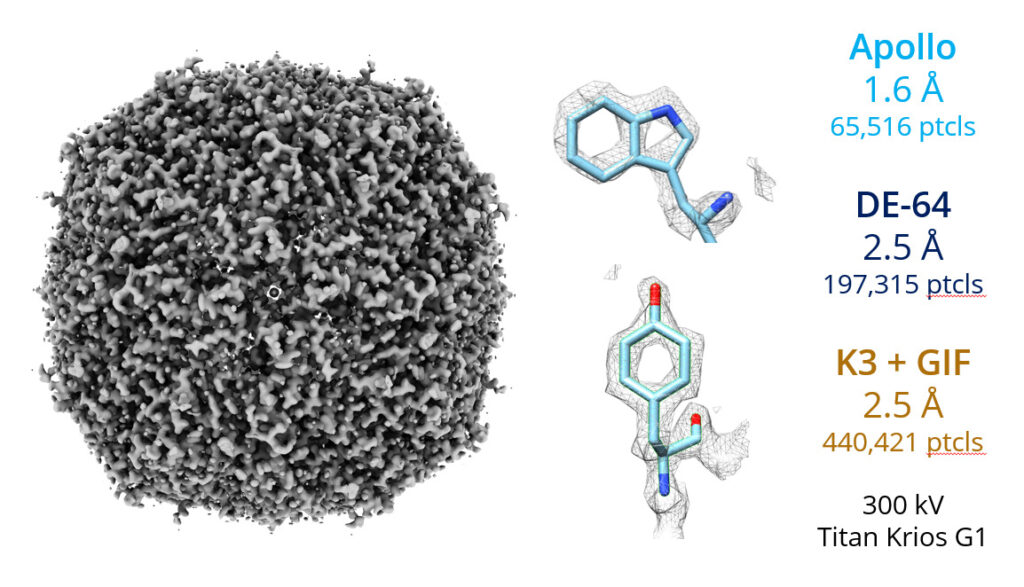
A recent paper from Florida State University has teased out the contributions of the number of particles used to find a protein structure versus the dose rate used. This work is of immense importance given that access to a Cryo-EM facility can cost ~$100/hr. “i.e optimizing the experimental conditions to increase throughput can save users tens or hundreds of thousands of dollars per year.”
Peng et al. prepared samples of the protein Apoferritin and performed single particle analysis using datasets collected at different dose rates on the same sample1. As expected, by adding successively more and more images, averaging together more and more apoferritin particles, the resolution of the protein structure was improved regardless of the dose rate. However, by noting the resolution of the structure achieved with each different number of particles, the researchers were able to graph out the benefit of ‘number of particles’ to the resolution of the structure. By replicating this dataset at different dose rates, they were able to disentangle the impact of ‘dose rate’ from ‘number of particles’ on the final resolution of the protein structure.
WHAT IS THE MAIN TAKEAWAY?
While it is not obvious visually, the authors note that the slope of each series is significantly different with 15 eps (electrons per pixel per second) having the steepest slope. This means that the information contributed by each additional particle is less at higher dose rates, specifically, 8% less at 30 eps and 15% less at 60 eps. Why? Because while each particle was given the same total dose (60 e−/Å2 ) detected on the camera, coincidence loss increases and causes more information to be lost at higher dose rates. Somewhat counterintuitively, while that means that the particles received a higher total dose at 60 eps, some fraction of the very first electrons to impact each particle (which contain the most pristine high frequency information), are not detected. This is what causes the flatter slope and reduced benefit of particle averaging at higher dose rates.
WHAT DOES THIS MEAN IS PRACTICE?
Firstly, the dose rate used should be chosen based on the desired resolution needed from the dataset. If the data is being acquired for sample screening, and high resolution is not needed, then it seems beneficial to use a high dose rate to quickly acquire enough particles to reach a medium/low resolution structure. However if a high resolution structure is desired the choice is more complicated. Take for example and initial apoferritin resolution of 2.33Å obtained using the conditions at point (i) in Figure 1, 3000 particles, 60 eps. If you wanted to achieve 2.04Å resolution instead, you could use the conditions at point (ii) and acquire 11,000 particles at 60 eps , or use the conditions at point (iii), 2500 particles at 15 eps. In this case the choice is essentially to acquire 4x slower or to acquire 4x more data, so both will take approximately the same amount of time. The only consideration that weighs in favor of point (iii) is that the data will take less space and can be processed more quickly.
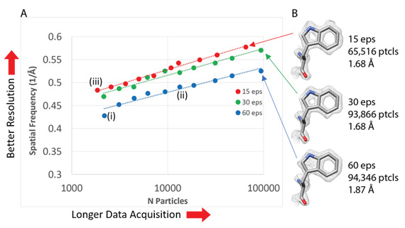
Figure 1: Adapted from Peng et al. (a) a graph of apoferritin refined structure resolution versus number of particles used, for various dose rates. (b) Tryptophan density maps of Apoferritin found for various dose rates.
By replicating this dataset at different dose rates, they were able to disentangle the impact of ‘dose rate’ from ‘number of particles’ on the final resolution of the protein structure.
To what extent can these results be applied to other experimental setups?
There will of course be experimental differences, but these results provide a valuable framework to understand which dose rate cryo-EM users should choose for their experiments.
References:
