
Single-particle cryo-EM is a proven technique for solving the structure of biological macromolecules, from membrane proteins to viruses and antibodies.
As such is has become a critical tool for structural biologists at universities and pharmaceutical companies worldwide. Single-particle-analysis (SPA) cryo-EM provides the key information in drug development, vaccine development and therapeutic treatments that protects humans against sickness and disease.
Electrons from a 300kV Cryo TEM produce a high resolution image of each protein crystal on the sample. However many cryo-EM cameras have 2 key characteristics that throttle the productivity and throughput for Cryo TEMs:
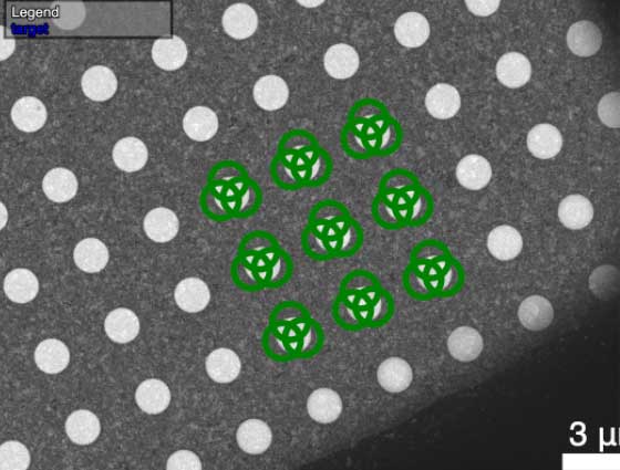
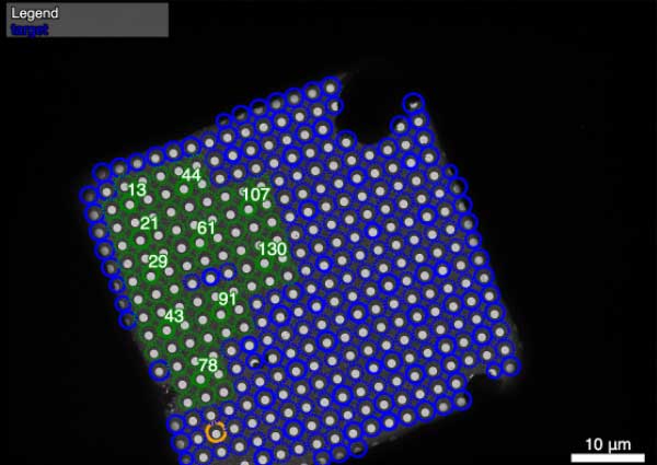
Figure 1: multi-shot per hole single particle data acquisition using SmartScope
1) MISPLACING ELECTRONS
At low spatial frequencies i.e. coarse features, a good counting detector localizes electrons accurately, so DQE is at its highest, set largely by how efficiently the detector counts electrons in the first place. The harder regime is high spatial frequency, near the detector’s Nyquist limit, where the finest structural detail lives. Here, any error in pinpointing each electron — charge shared between adjacent pixels, or imprecise centroiding — blurs fine detail and pulls DQE down.
A critical ingredient for keeping DQE high near Nyquist is therefore the physical pixel size. Relative to a detector’s own Nyquist limit, larger pixels contain each electron’s deposited signal more completely than small pixels, reducing blurring and preserving high-resolution detail. The physical pixel sizes of the most popular detectors are shown below.
Both the Apollo and the K3 recover DQE by outputting super-resolution images, where the charge is localized to a quadrant within a pixel, to further improve DQE near Nyquist. They also use CDS (a noise reduction technique) to enhance SNR and DQE.
| CAMERA | FALCON4i | APOLLO | K3 |
|---|---|---|---|
|
Pixel Size: |
14µm |
8µm |
5µm |
Both the Apollo and the K3 recover DQE by outputting super-resolution images, where the charge is localized to a quadrant within a pixel, to further improve DQE near Nyquist. They also use CDS (a noise reduction technique) to enhance SNR and DQE.
2) RECORDING ELECTRONS TOO SLOWLY
When two electrons arrive at the same pixel in a frame, or in nearby pixels where their charge is connected, counting algorithms consider them to be one electron that just deposited a lot of energy. This results in one of the electrons being erased completely from the image, and is known as coincidence loss. Coincidence loss degrades DQE at high dose rate → to protect DQE you must cap the dose rate → the capped dose rate forces longer exposures → longer exposures cap throughput.
The key criteria for co-incidence loss to occur is that the electrons must arrive in the same frame. Increasing the camera framerate reduces coincidence loss, maintaining high DQE at high dose rates, and thus unlocking high throughput.
Most labs carry out single-particle-analysis on a Falcon4i using a dose rate of 4-5 e/pix/s. 4.6 e/pix/s corresponds to a 10% drop in DQE due to co-incidence loss, so here we compare the speed of each cryo-EM camera under the same 10% coincidence loss limitation.
| CAMERA | FALCON4i 1 | APOLLO 3 | K3 2 |
|---|---|---|---|
|
Internal framerate: |
320 fps (no CDS) |
2,620 fps (w/ CDS) |
750 fps (W/ CDS) |
|
Image shift via Low Dose Mode (LDM) |
4.6 e/pix/s |
33.0 e/pix/s |
8.3 e/pix/s |
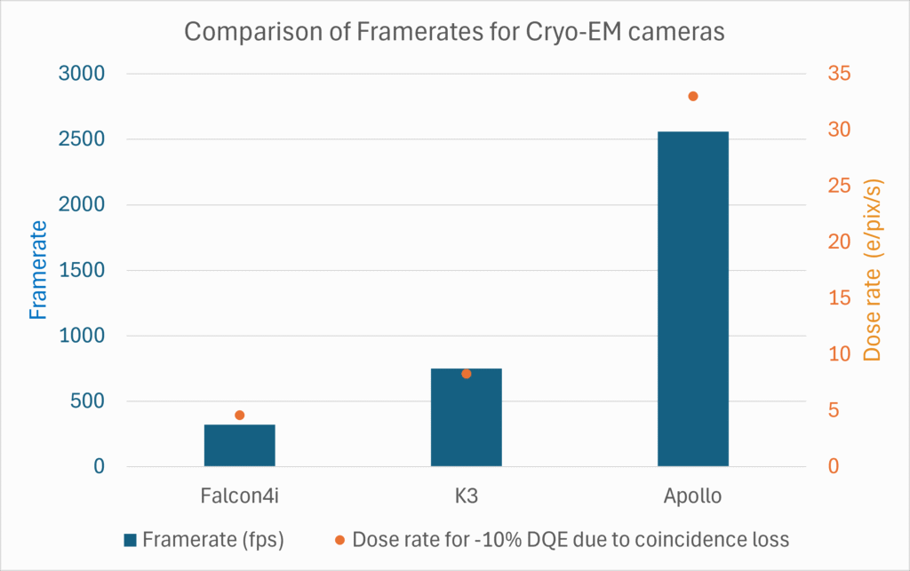
The Falcon4i is ~7x slower than Apollo, while the K3 is ~4x slower than Apollo. Thus Apollo can operate at a much higher dose rate with the same coincidence loss,
Both the Falcon4i and Apollo augment their framerate by outputting dose-fractionated, event-streaming data formats (EER and CSB respectively). This allows users to isolate the first few electrons to impact a protein and upweight/prioritize that high resolution data before the structure has been damaged.
SUMMARY
Overall, the strength of the Falcon4i is it’s large pixel size and EER data format, while it’s drawbacks are a much slower counting speed and lack of CDS. The strength of the K3 is it’s CDS and super-resolution output, while it’s drawbacks are a slower counting speed than Apollo, small pixel size and lack of event-streaming data format. Apollo combines a good pixel size and the fastest counting speed, with all the benefits of CDS, event-streaming data format, and super resolution readout.
HOW DOES CAMERA SPEED TRANSLATE TO SINGLE PARTICLE THROUGHPUT?
Single-particle-analysis works by recording many movies of an area of dispersed protein particles. To achieve a high resolution structure, a set number of movies must be collected e.g. 5000. With modern Cryo-TEMs such as the TFS Krios or the JEOL Cryo-ARM, fringe-free imaging means that image shift can be used to collect a series of movies, with ~0.5s delay between movies. Then stage shift must be used to move to a new area for next series of movies. There is a delay of ~2s or more after each stage shift.
Depending on the magnification and type of grid used, the use of image shift can be maximized relative to stage shifts to reduce overall delay. Overhead for acquiring and saving each movie varies depending on the camera and software used. This camera delay is in the range of 0.4s – 1.2s
Finally, there are additional delays due to microscope procedures such as re-focusing, sample tracking, initial grid montaging, sample changes, and liquid nitrogen refills.
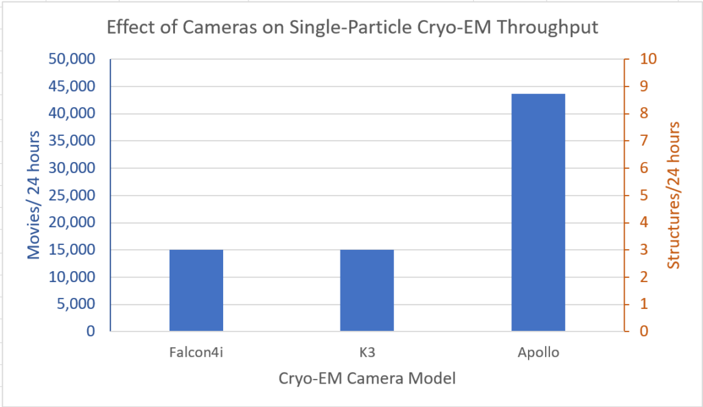
Overall, in real-world conditions, the throughput on high-end cryo-TEMs equipped with different cameras works out as follows:
CONCLUSION:
As improvements in stage movements, grid preparation and image-shift reduce the delay between movie acquisitions, camera speed has become a major limiting factor in cryo-em throughput. By switching from a K3 or Falcon4i, to the faster Apollo, facilities can almost triple their single-particle-analysis throughput. This can save facilities millions of dollars by obsoleting the need to acquire multiple microscopes.
References:
