DIRECT ELECTRON INTRODUCES CELERITAS
THE WORLD’S FASTEST 4D STEM CAMERA

San Diego, CA (USA), June 30, 2021 – Direct Electron, a leading producer of next-generation direct detection cameras for transmission electron microscopes (TEM), today announces Celeritas, a revolutionary new direct detection camera for four-dimensional scanning transmission electron microscopy (4D STEM). Based on a next-generation direct detection sensor, Celeritas far exceeds the frame rate of other 4D STEM detectors while also delivering at least four times more pixels and exceptional image quality.
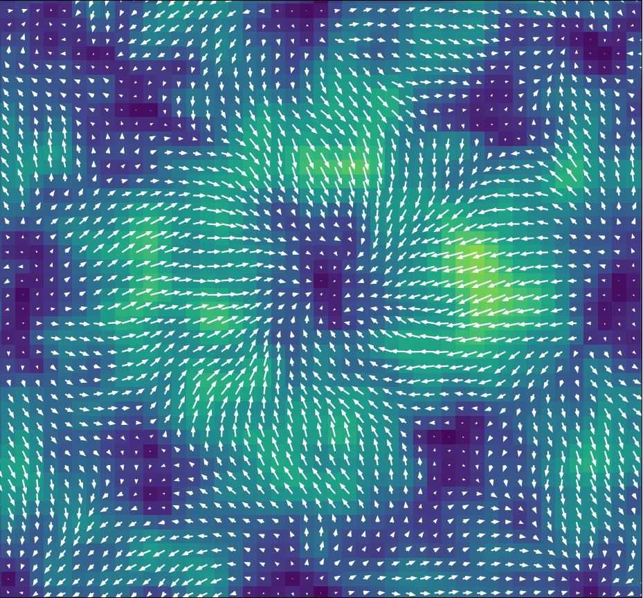
4D STEM is a cutting-edge technique that uses the richness of information available in STEM for exploring the structure of materials. Rather than a single averaged signal, 4D STEM cameras capture the full electron diffraction pattern so that researchers can peer into the subtle details of data, visualizing atoms that are difficult to see in conventional STEM and extracting information about crystal orientation, strain, electric and magnetic fields, and other important features.
However, until now researchers have had limited choices for 4D STEM detectors. Previous generation 4D STEM detectors took about 15-20 minutes to capture a one-megapixel STEM image, which is far too slow to be practical.
Celeritas is the Latin word for speed, and we could not think of a better word to describe a camera with acquisition speeds up to 100x faster, enabling a one-megapixel STEM image to be captured in about 10 seconds. This introduces a paradigm-shift for 4D STEM, enabling users, for the first time, to capture 4D STEM datasets at speeds comparable to conventional STEM images. 4D STEM experiments that would require hours of data collection with another detector can be performed in minutes using Celeritas.
In addition to its speed, a high dynamic range (HDR) imaging mode offers over 10x higher dynamic range per frame than was achievable in previous monolithic active pixel sensor direct detectors. Fast frame rates and a large number of pixels also facilitate greater dynamic range and enable higher probe currents to be used to acquire data, giving Celeritas the flexibility to operate over an extremely wide range of imaging conditions.
With one million (1024 x 1024) pixels, Celeritas is also an excellent high-speed TEM camera, enabling applications such as ultrafast, sub-millisecond time-resolution in situ TEM imaging.
The advances in both the range of imaging conditions and data throughput offered by Celeritas promise to profoundly increase the productivity of TEM facilities and accelerate discovery in materials sciences.
Dr. Benjamin Bammes, Director of Research and Development at Direct Electron, said, “The speed of Celeritas is remarkable, particularly because we managed to achieve its ultrafast frame rate without sacrificing image quality, pixel area, or dynamic range. This has been a major development effort at Direct Electron over the past five years, building on our lengthy experience in designing advanced low-noise direct detectors and driven by the demands of the materials science community that is always pushing the boundaries of technology.”
Throughout its development, materials science experts provided feedback and advice for Celeritas. These include Prof. Paul Voyles at the Wisconsin Materials Research Science and Engineering Center at the University of Wisconsin-Madison, and Prof. Angus Kirkland of Oxford University and the Rosalind Franklin Institute in the United Kingdom. The first Celeritas camera was installed in the Nanoscale Imaging and Analysis Center directed by Prof. Voyles, and the second Celeritas camera will be installed at the Rosalind Franklin Institute under the direction of Prof. Kirkland.
Prof. Paul Voyles said, “I’m tremendously excited about the Celeritas camera. The high speed lets us acquire 4D STEM data like strain maps over large areas and outrun some of the instrumental instabilities of our microscope. It also enables a variety of time-resolved in situ 4D STEM experiments that are impossible without it, like measuring crystal nucleation and growth rates and characterizing the fluctuating atomic structure of supercooled liquids.”
Celeritas is a complete system that includes a high-performance computer and software in addition to the camera module. It is compatible with most commercial TEM/STEM microscopes.
Celeritas is available to order immediately, with delivery for orders placed now expected within the fourth quarter (Q4) of this year.