DE CAMERA USED IN STRUCTURAL BIOLOGY PAPER OF THE YEAR

Congratulations to Axel Brilot and colleagues on being awarded the Journal of Structural Biology Paper of the Year.

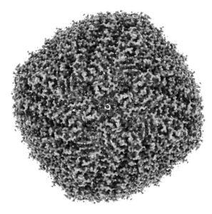
One of the principle investigators of the paper, Nikolaus Grigorieff, summarized it as follows: “Beam-induced motion, first described by Henderson and Glaeser in 1985, is a well-known problem that has plagued users of electron cryo-microscopy (cryo-EM) for decades. The motion occurs while the electron beam irradiates the sample and leads to significant blurring of the image and, ultimately, to loss of resolution in three-dimensional reconstructions of the visualized molecules.” In order to study beam-induced motion, Brilot and colleagues used the first commercially-available direct detector for TEM: a Direct Electron DE-12 Camera System. Dr. Grigorieff continued: “In addition to the higher sensitivity (DQE) compared to older detectors, the CMOS technology underlying the DE-12 camera is also able to record movies at a rate of 40 frames/second, ideal for the study of motion under the beam. Jointly with the Carragher/Potter lab, Axel and Anchi Cheng continued the study of beam-induced motion of DLPs using the DE-12 camera. They were the first to demonstrate that image blurring produced by beaminduced motion on ice-embedded specimens can be significantly reduced by aligning movie frames. This post-processing of electron micrographs thus overcomes one of the major obstacles limiting the resolution of cryo-EM structures. Movies have since transformed the field of cryo-EM and are now in common use.”

Dr. Briolot said that collaboration with Direct Electron critical during this study: “What I found most amazing about working with Direct Electron was how responsive they were in working with Anchi Cheng and I to implement movie mode acquisition and enable us to visualize and correct beam-induced motion. I was amazed at the time, but am even more so now, realizing that DE and Anchi were able to implement ‘movie mode’ in what seemed like just hours, with almost no hiccups once we started working.”

Categories

What’s New

Read More

Dr. Barnaby Levin to Present at MSC-SMC 2026 in Montreal

Direct Electron is pleased to announce that Dr. Barnaby Levin will be presenting at the 52nd Annual Meeting of the Microscopy Society of Canada / Société de Microscopie du Canada (MSC-SMC) in Montreal. MSC-SMC 2026 brings together researchers and technologists from across the microscopy community for a week of scientific exchange focused on electron microscopy, advanced imaging, analytical techniques, and emerging instrumentation. The meeting will also feature a Microscopy Summer School on May 25–26, including lectures and hands-on sessions in analytical electron microscopy, imaging data processing, advanced optical microscopy, and X-ray microscopy. We look forward to connecting with attendees throughout the meeting to discuss the latest advances in cryo-EM, diffraction, and high-performance direct detectors for TEM imaging. If you’ll be attending MSC-SMC 2026 in Montreal, we hope you’ll join Dr. Barnaby Levin’s presentation and connect with our Canadian partners Edge Scientific during the meeting. And don’t forget to follow Barnaby on BlueSky at @bdalev.bsky.social!

Dr. Barnaby Levin Attending TEM Gordon Research Conference

We’re pleased to share that our Applications Scientist, Dr. Barnaby Levin, is attending the upcoming Transmission Electron Microscopy for Materials Research Gordon Research Conference, one of the premier international meetings focused on advancing the frontiers of TEM science and instrumentation, to give a talk about cutting-edge MAPS Detector Technology. The conference, running February 15-20, brings together leading researchers from around the world to present cutting-edge, often unpublished work while fostering deep scientific discussion and collaboration. This year’s program highlights recent breakthroughs in high-resolution TEM imaging and spectroscopy, while addressing key challenges such as imaging radiation-sensitive materials, improving throughput, and resolving structures in thicker samples. As TEM technology continues to push toward theoretical limits, meetings like this help shape the next decade of innovation in materials characterization. We’re excited to see Barnaby engaging with the global TEM community and contributing to discussions driving the field forward. And don’t forget to follow him on BlueSky at @bdalev.bsky.social!

Questions?